Among the earliest inputs that cells experienced, mechanical stress (forces) guide and direct behavior of cells, including when they are part of tissues, organs, and organ systems. These mechanical stresses are propagated through the cell’s skin (the cell cortex), which is a composite material of membrane and cytoskeleton. Key molecular machinery senses the forces, and through mechanotransduction, the mechanical signals may be converted into biochemical signals, which guide cell behavior.
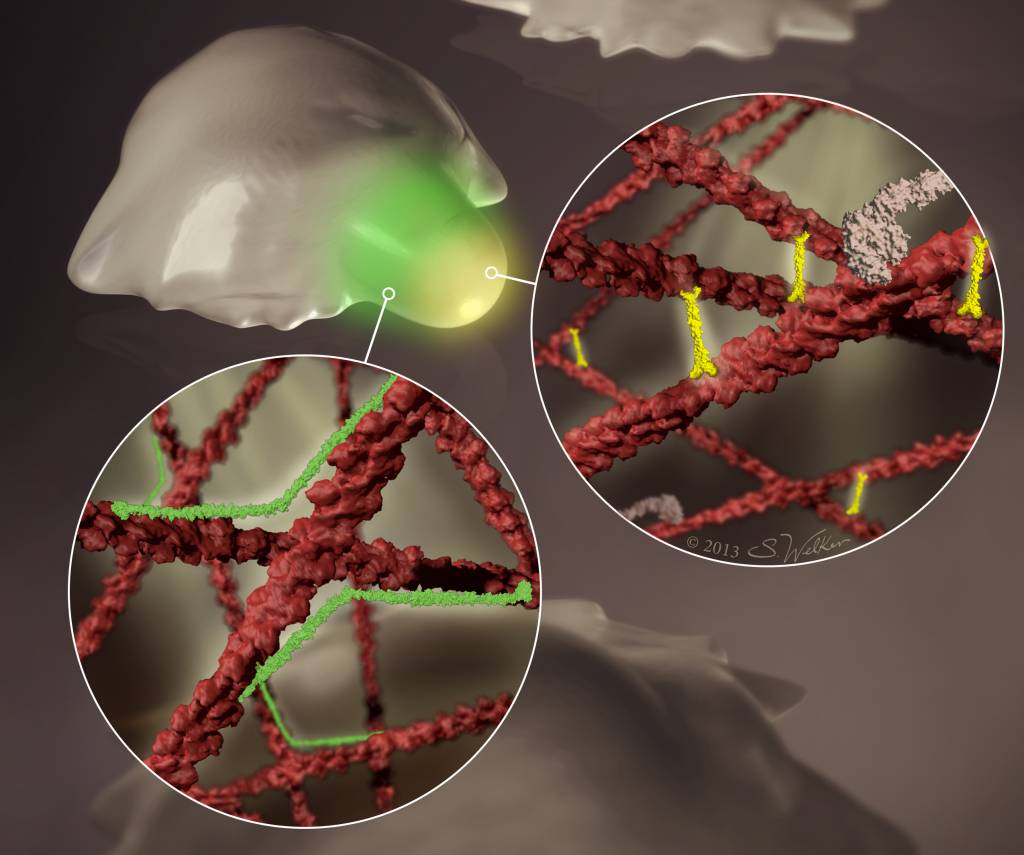
This image depicts a cell experiencing mechanical stress, which imposes dilation and shear deformations. Different molecular machinery accumulates in response to each type of deformation. The region highlighted in green experiences shear deformation. The inset shows that the actin crosslinking protein filamin (green) binds to the crossed actin filaments (red), allowing filamin to sense shear deformation. The yellow region experiences dilation deformation. In this case, the inset shows that a different set of proteins, including the mechanoenzyme myosin II (white) and the actin crosslinker α-actinin, sense and accumulate in response to this type of deformation. These protein accumulations may now be explained quantitatively based on each protein’s molecular mechanisms. These protein accumulations, along with the cell’s viscoelastic makeup, account for how nonmuscle cells contract against mechanical stress.
The art work was kindly prepared by Art as Applied to Medicine graduate student, Samantha Welker.